ADVANCED ANALYSIS FOR SPIROMETRY
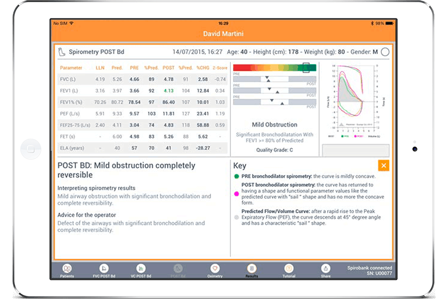
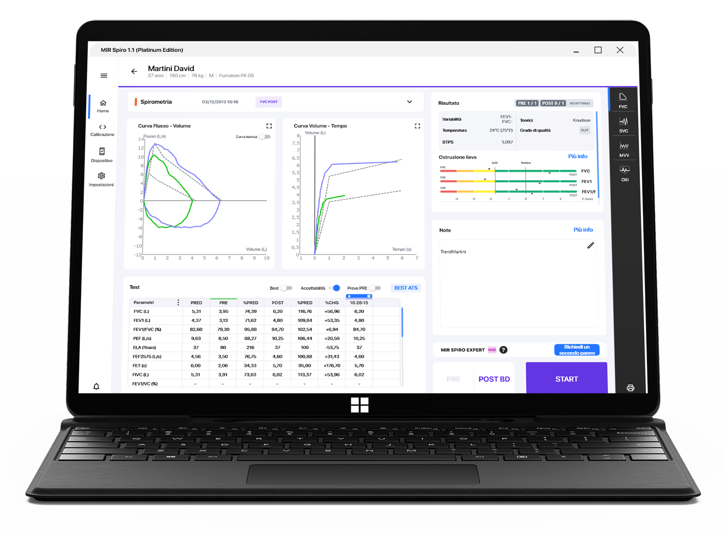
Session summary with FVC, SVC, MVV; FVC History for session comparisons.
Editing tools to:
- Set Best trial
- Disable/enable/delete/recover trials
- Configure parameters to display and in what order
: The video library spans almost every chapter, covering a vast range of specialties from whole-body imaging to specialized fetal, obstetric, and pediatric sonography. Why Rumack Ultrasound Videos are Essential
In short, if the textbook is your anatomy atlas, the video library is your clinical rotation.
Videos cover everything from pediatric imaging to complex image-guided procedures . How to Access the Content
Real-world examples of deep vein thrombosis (DVT) compression tests. 2. Abdominal and Small Parts Imaging
Whenever an instructional video features a picture-in-picture view of the sonographer's hand, pay close attention. Note how the probe is oriented relative to the patient’s anatomy and how pressure is applied to optimize the acoustic window. Step 3: Test Your Recognition Skills
Medical institutions utilize video libraries to standardize scanning protocols across large hospital systems. By aligning staff competencies with the video criteria established in standard reference materials, departments can minimize inter-operator variability, ensuring that a scan performed by a night-shift technician maintains the same diagnostic integrity as one performed by a senior department head. Enhanced Remediation and Self-Directed Study
The library spans several critical areas, including:
: Fetal development stages, anatomy scans, and screenings for anomalies.
The videos are shot using modern ultrasound equipment with high frame rates and clear annotation. Subtle findings—like the "twinkling artifact" behind a calculus or the "yin-yang sign" of a pseudoaneurysm—are unmistakable.
Each video is cross-referenced to specific chapters and figures in Diagnostic Ultrasound . You can read about the sonographic appearance of a liver hemangioma and then immediately watch a video showing its characteristic peripheral nodular enhancement on contrast ultrasound.
Demonstrating the definitive diagnostic maneuver for DVT—complete venous compressibility under direct manual pressure.
Unlocking the Gold Standard: A Guide to the Rumack Ultrasound Video Library
While textbooks offer foundational knowledge, ultrasound is a dynamic, real-time modality. This is why have become indispensable tools for sonographers, radiologists, and medical students worldwide. Watching a live scan provides spatial and temporal context that a flat textbook image simply cannot match. Why Rumack Ultrasound Videos are Essential for Learning
are the premier real-time companion clips to the industry-standard textbook Diagnostic Ultrasound , edited by world-renowned experts Dr. Carol M. Rumack and Dr. Deborah Levine . These multimedia assets bridge the gap between static print and actual clinical scanning, serving as an essential masterclass for radiologists, sonographers, and medical residents. Across modern editions like the sixth edition published by Elsevier , the textbook features approximately 400 video clips . These clips capture live anatomic movement, hemodynamic flow patterns, and real-time pathologies across every human organ system. What are Rumack Ultrasound Videos?
Before diving into the video content, it is important to understand the authority behind the name. Carol M. Rumack, MD, FACR, is a Professor of Radiology and Pediatrics at the University of Colorado School of Medicine. She was and continues to practice neonatal imaging in the high-risk neonatal intensive care unit (NICU). Her research has centered on neonatal sonography of high-risk infants, particularly the brain.
Eliminating wasted space at the bottom of the screen to increase frame rate. How to Integrate Video Learning into Your CME Routine
Note: Be cautious of "free" Rumack videos on dubious websites. These are often low-resolution, outdated (from the 3rd or 4th edition), or violate copyright. Investing in the official resource supports continued updates.
: Videos are typically accessed via an eBook included with the print purchase, allowing for integrated learning on mobile devices or computers. Key Clinical Topics Covered
Session summary with FVC, SVC, MVV; FVC History for session comparisons.
Editing tools to:
- Set Best trial
- Disable/enable/delete/recover trials
- Configure parameters to display and in what order
Specific analysis application:
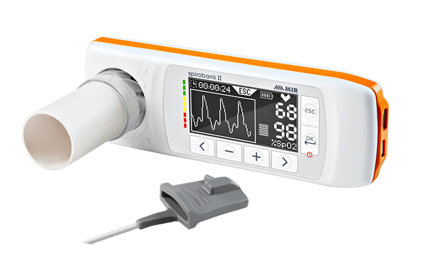
- 6-Minute Walk Test (6MWT)
- Sleep Test
- 24-hour Holter saturation with adjustable titration
Architecture strongly oriented towards interoperability optimizing workflows and data exchange with EMR/EHR. Numerous standards supported such as HL7, FHIR (Json), GDT, DICOM, eXchange Protocol, and many others.
Patient list, printing, data export.
Support up to 22 languages.
Real-time animation to improve patient collaboration during the test. Based on an algorithm that takes into account both Flow and Volume to make it more reliable and effective.
ATS2019, Winspiro classic, NIOSH, OSHA.
Import of tests from MIR professional devices.
Access all the benefits offered by MIR Spiro, enjoy your Platinum experience!
Exchange data without limits between MIR Spiro and external platforms
Be amazed by innovation. Keep up with the latest trends
Get live support from a MIR operator wherever and whenever you need. Includes 1 free session of remote video assistance
One single database, multiple devices. A shared database for all workstations on the same local network, designed for clinics, medical centers, and healthcare facilities.
Start now your
Platinum experience
With your Platinum subscription plan, you will have uninterrupted access to all features of MIR Spiro, exchange data unlimitedly and free of charge between MIR Spiro and remote platforms, and access extra content while staying updated on the latest trends, all without limits!
Additionally, you will have access to free technical support from a MIR operator ready to assist you wherever and whenever you need. 1 remote technical assistance session is included.
Experience the best, choose MIR Spiro Platinum.
ADVANCED SPIROMETRY TREND
For each patient, the user can select a parameter and check its trend over the selected time period.
FREE ACCESS TO VIDEO TUTORIALS
Exclusive to subscribers, unlimited access to video tutorials on software and device usage.
BIDIRECTIONAL WORK LIST
Data exchange has never been easier! Create your patient list on MIR Spiro and send it with a click to your MIR device. Perform the test with the device in Stand Alone mode and import the results into MIR Spiro.
Chinese (China), Chinese (Taiwan), Czech (Czechia), Dutch (Netherlands), English (United Kingdom), English (United States), French (France), French (Belgium), Georgian (Georgia), German (Germany), Hungarian (Hungary), Italian (Italy), Japanese (Japan), Latvian (Latvia), Polish (Poland), Portuguese (Portugal), Romanian (Romania), Russian (Russia), Spanish (Spain), Swedish (Sweden), Turkish (Turkey), Ukrainian (Ukraine)
WINDOWS
MACOS
: The video library spans almost every chapter, covering a vast range of specialties from whole-body imaging to specialized fetal, obstetric, and pediatric sonography. Why Rumack Ultrasound Videos are Essential
In short, if the textbook is your anatomy atlas, the video library is your clinical rotation.
Videos cover everything from pediatric imaging to complex image-guided procedures . How to Access the Content
Real-world examples of deep vein thrombosis (DVT) compression tests. 2. Abdominal and Small Parts Imaging
Whenever an instructional video features a picture-in-picture view of the sonographer's hand, pay close attention. Note how the probe is oriented relative to the patient’s anatomy and how pressure is applied to optimize the acoustic window. Step 3: Test Your Recognition Skills
Medical institutions utilize video libraries to standardize scanning protocols across large hospital systems. By aligning staff competencies with the video criteria established in standard reference materials, departments can minimize inter-operator variability, ensuring that a scan performed by a night-shift technician maintains the same diagnostic integrity as one performed by a senior department head. Enhanced Remediation and Self-Directed Study
The library spans several critical areas, including:
: Fetal development stages, anatomy scans, and screenings for anomalies.
The videos are shot using modern ultrasound equipment with high frame rates and clear annotation. Subtle findings—like the "twinkling artifact" behind a calculus or the "yin-yang sign" of a pseudoaneurysm—are unmistakable.
Each video is cross-referenced to specific chapters and figures in Diagnostic Ultrasound . You can read about the sonographic appearance of a liver hemangioma and then immediately watch a video showing its characteristic peripheral nodular enhancement on contrast ultrasound.
Demonstrating the definitive diagnostic maneuver for DVT—complete venous compressibility under direct manual pressure.
Unlocking the Gold Standard: A Guide to the Rumack Ultrasound Video Library
While textbooks offer foundational knowledge, ultrasound is a dynamic, real-time modality. This is why have become indispensable tools for sonographers, radiologists, and medical students worldwide. Watching a live scan provides spatial and temporal context that a flat textbook image simply cannot match. Why Rumack Ultrasound Videos are Essential for Learning
are the premier real-time companion clips to the industry-standard textbook Diagnostic Ultrasound , edited by world-renowned experts Dr. Carol M. Rumack and Dr. Deborah Levine . These multimedia assets bridge the gap between static print and actual clinical scanning, serving as an essential masterclass for radiologists, sonographers, and medical residents. Across modern editions like the sixth edition published by Elsevier , the textbook features approximately 400 video clips . These clips capture live anatomic movement, hemodynamic flow patterns, and real-time pathologies across every human organ system. What are Rumack Ultrasound Videos?
Before diving into the video content, it is important to understand the authority behind the name. Carol M. Rumack, MD, FACR, is a Professor of Radiology and Pediatrics at the University of Colorado School of Medicine. She was and continues to practice neonatal imaging in the high-risk neonatal intensive care unit (NICU). Her research has centered on neonatal sonography of high-risk infants, particularly the brain.
Eliminating wasted space at the bottom of the screen to increase frame rate. How to Integrate Video Learning into Your CME Routine
Note: Be cautious of "free" Rumack videos on dubious websites. These are often low-resolution, outdated (from the 3rd or 4th edition), or violate copyright. Investing in the official resource supports continued updates.
: Videos are typically accessed via an eBook included with the print purchase, allowing for integrated learning on mobile devices or computers. Key Clinical Topics Covered

State accedendo ad un’area che contiene informazioni dettagliate di carattere promozionale sui prodotti della società rivolte esclusivamente agli operatori professionali e pertanto non fruibili da soggetti non qualificati (art. 21 D.Lgs. 46/97).
Ai sensi del decreto legislativo n. 46/1997, e del decreto del Ministero della Salute del 23 febbraio 2006, e successive modifiche intervenute, Vi preghiamo, se siete Professionisti Sanitari, di cliccare sul pulsante "Consenti" per accedere.